Tag: diagnostic imaging

  • Shoulder MRI Scan: What It Shows and What to Expect

    Shoulder MRI Scan: What It Shows and What to Expect

    Could the source of shoulder pain be in structures that an X-ray cannot show clearly? When symptoms persist or an injury needs closer assessment, a clinician may recommend a comprehensive shoulder MRI scan to examine soft tissues and other structures in detail. You may be wondering why MRI is the next step, what it could reveal, or whether staying still in the scanner will feel uncomfortable.

    This guide explains why a clinician may recommend a shoulder MRI and what the images can help assess, including tendons, ligaments, cartilage and surrounding tissues. You’ll also learn what to share before the scan, how to prepare and what happens during imaging. If enclosed spaces make you uneasy, a specialized scan at Open MRI Dubai may offer a more comfortable environment. Your referral and the clinical question guide the imaging plan.

    After the scan, a radiologist interprets the images and prepares a report for your referring clinician. Your clinician discusses the findings alongside your symptoms and examination. Knowing how these steps fit together can help you feel more prepared for your appointment.

    Key Takeaways

    • A clinician recommends a shoulder MRI based on your symptoms, examination, medical history and the question the scan needs to answer.
    • MRI uses magnetic fields and radiofrequency signals to create views of structures inside and around the shoulder joint.
    • Open-format scanners have a different physical design that may feel more comfortable for some people. The imaging plan still depends on the clinical need.
    • Share relevant medical history, implant information, previous imaging and referral details with the imaging team, and follow their preparation instructions.
    • If you’re considering a shoulder mri dubai scan, remember that the radiologist’s report and your clinician’s assessment work together to guide next steps.

    Why might a clinician recommend a shoulder MRI scan?

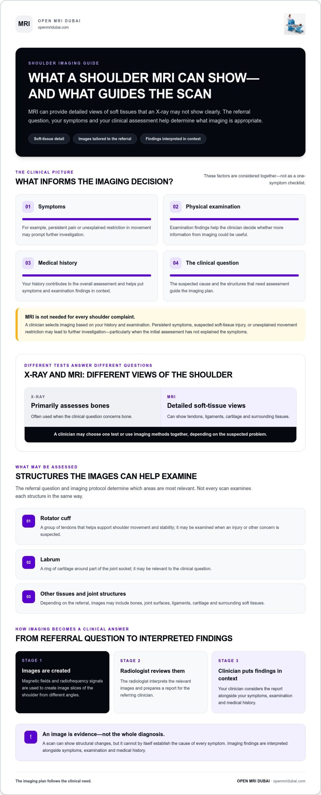
    A shoulder MRI, or magnetic resonance imaging scan, uses magnetic fields and radiofrequency signals to create detailed images of structures inside and around the joint. An X-ray is primarily used to assess bones, while MRI can provide views of soft tissues such as tendons, ligaments and cartilage. A clinician may recommend MRI when images of those structures could help answer a specific question about your shoulder.

    The decision depends on the full clinical picture: your symptoms, physical examination, medical history and the suspected cause of the problem. Persistent pain, a suspected soft-tissue injury or unexplained restriction in movement may prompt further investigation, particularly if the initial assessment hasn’t explained your symptoms. Not every shoulder complaint requires MRI. The referring clinician selects imaging based on what they need to assess.

    For people researching shoulder mri dubai, understanding the purpose of the scan can make a referral feel less uncertain. MRI is one part of an investigation, not a replacement for a consultation or examination.

    What questions can a shoulder MRI help investigate?

    Depending on the clinical question and imaging protocol, a radiologist may assess the shoulder’s tendons, ligaments, cartilage and other soft tissues. The rotator cuff, a group of tendons that helps support shoulder movement, may be examined when an injury or another concern is suspected. The labrum, a ring of cartilage around part of the joint, may also be relevant.

    For example, a clinician might request MRI to investigate ongoing pain or reduced movement when they need more information about soft tissues than an X-ray can provide. Conditions such as shoulder impingement syndrome can be associated with shoulder pain, but symptoms and imaging findings need careful interpretation. A scan can show structural changes; it cannot, by itself, establish the cause of every symptom.

    When might MRI be considered alongside other imaging?

    Different imaging tests answer different questions. X-rays are commonly used to assess bones, while MRI can offer detailed views of soft tissues. Depending on the suspected problem, a clinician may choose one test or use imaging methods together. The appropriate choice depends on your history and examination, not on a single symptom or a general preference for one scan type.

    An imaging test provides evidence about structures. A complete clinical diagnosis brings that evidence together with symptoms, examination and medical history. A finding on an image may not fully explain how your shoulder feels or moves. Your referring clinician interprets the radiology report in context and explains what the findings mean for your care.

    How does a shoulder MRI create detailed images of the joint?

    An MRI scanner uses a strong magnetic field and radiofrequency signals to create images of the body. In simple terms, these signals interact with tissues in different ways, and the scanner records the responses. A computer processes that information into image slices, giving the radiologist views through the shoulder from different angles.

    These views let the radiologist examine the area relevant to the referral rather than relying on a single flat picture. The shoulder includes the glenohumeral joint, where the rounded top of the humerus, or upper arm bone, meets the socket of the shoulder blade. The scan can show this joint and nearby tissues. The precise images and sequences are selected according to the referral question and clinical judgment.

    Which shoulder structures may appear in the images?

    The scan may include bones, joint surfaces and surrounding soft tissues. The rotator cuff is a group of tendons that supports shoulder movement and stability. The labrum is a ring of cartilage around the joint socket. It may be assessed when relevant to the clinical question, but not every scan examines each structure in the same way. The radiologist reviews the images in light of the requested assessment.

    If you’re learning about shoulder mri dubai, it helps to know that the scan isn’t simply a general picture of the whole shoulder. Its image views are planned to address the reason for imaging. For a patient-friendly overview of the scan and its role, see UCSF Health’s Shoulder MRI Scan: What It Shows and What to Expect.

    What does AI-assisted imaging mean for patients?

    Open MRI Dubai provides AI-powered diagnostic imaging, including open MRI scans. AI-powered imaging is part of the imaging service, while the images still require professional review. It does not replace a radiologist’s interpretation or a clinician’s assessment of what the findings mean for an individual patient.

    Imaging technology helps produce images; clinical interpretation puts those images in context. The radiologist reviews the relevant views and prepares a report. The referring clinician relates the findings to your symptoms and examination. Professional interpretation remains central to understanding the images.

    Knowing that image views are tailored to the clinical question can make the process feel more purposeful. If you’re exploring an open MRI scan, learning about the open-format approach can help you understand the imaging environment as well as the technology used to create the images.

    Open-format shoulder MRI: comfort benefits and practical considerations

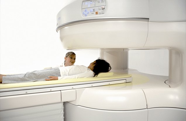
    An open-format MRI has a more open physical design than an enclosed scanner, which may feel less confining for some patients. This can be reassuring if enclosed spaces make you anxious or you simply prefer a more open environment. Comfort is personal, however. The scanner’s design is only one part of the experience and cannot guarantee that every person will feel relaxed during an examination.

    Open MRI Dubai provides open-format imaging, including sitting open MRI technology. The setup for a shoulder examination depends on the clinical question and imaging protocol. A scanner’s appearance alone doesn’t establish whether it can meet the needs of a particular examination or whether it is suitable for a specific patient.

    What can an open MRI environment feel like?

    Rather than being surrounded by a narrow tunnel, patients may have a more open space around them. That difference can make the scan feel more manageable for some people who dislike enclosed areas. The shoulder still needs to be positioned for the requested images, and you’ll need to follow the imaging team’s instructions and remain as still as you comfortably can while images are captured.

    People respond differently to the scanner environment, positioning and sounds during imaging. If you feel uneasy, tell the imaging team so they can explain what to expect and guide you through the process. An open design may support comfort, but it doesn’t remove the need for careful positioning or make the experience identical for every patient.

    How should patients think about scan suitability?

    Suitability depends on the body area being examined, the clinical question and the protocol selected by the imaging team. For a shoulder MRI, the requested assessment helps determine which images are needed. The scanner format alone doesn’t establish whether the examination will answer that question. Follow the instructions for your scan and share any relevant concerns or information that may affect positioning.

    If you’re comparing options for shoulder mri dubai, consider more than whether a scanner looks open or enclosed. The purpose of the examination and its imaging requirements matter just as much as the physical environment. A format that feels appealing may not suit every examination, and open MRI shouldn’t be assumed to produce the same images for every clinical need.

    For a broader look at the open-format experience and comfort considerations, explore this complete guide to open MRI imaging and patient comfort. It explains the environment; your referral and imaging instructions remain the guide for your individual examination.
    Shoulder MRI Scan: What It Shows and What to Expect

    How to prepare for a shoulder MRI scan and what happens during it

    A little preparation can make your appointment more straightforward. Follow the imaging centre’s specific instructions, as preparation and scan protocols can vary. Share relevant health information before the scan, especially details about implants or previous procedures. This helps the imaging team complete safety screening and plan the examination.

    Use this sequence as a guide, while treating the instructions for your own examination as the priority:

    1. Gather your documents. Bring your referral information and any previous shoulder images or reports that are available and relevant.
    2. Tell the team about your health history. Mention implants, medical devices, past procedures and health conditions that may affect your scan.
    3. Choose suitable clothing. Follow the centre’s directions about clothing and metal. You may be asked to change and remove metallic items, such as jewellery or a watch, before imaging.
    4. Ask about anything unclear. Raise questions about preparation, positioning or what to expect with the imaging team before the scan begins.

    What should you tell the imaging team beforehand?

    Give specific information about any implant or device, and share relevant details about previous procedures or medical conditions. If you have earlier shoulder imaging, bring it when available so the team has useful context. Your referral information also helps identify the clinical question behind the examination. Metal screening is a routine MRI safety step; accurate information helps staff plan the imaging appropriately.

    What happens while the images are taken?

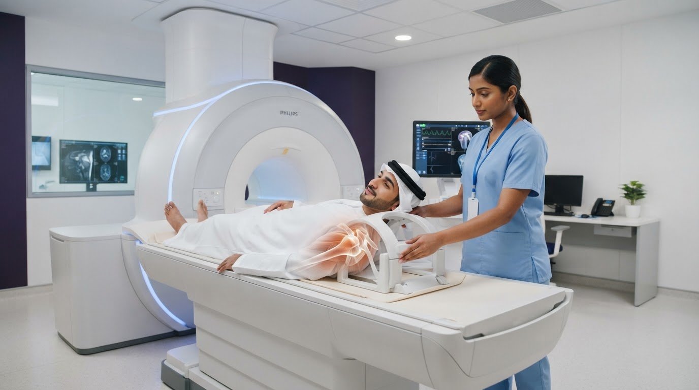
    The imaging team will guide you into a position for the shoulder examination and explain how to stay still while images are captured. Careful positioning helps produce useful images. The scan may include multiple image sequences, with the protocol shaped by the referral question and clinical judgment. The team will guide you through the process, so you don’t need to know the sequence in advance.

    Staying still can be challenging if your shoulder is uncomfortable. Tell the staff if you’re struggling to maintain the position or feel uneasy. They can guide you and explain what they need you to do. Avoid changing position without instructions, as movement may affect the images and a sequence may need to be repeated.

    If you’re preparing for a shoulder mri dubai appointment, following your specific instructions and sharing relevant information can help the team plan the examination. Explore open MRI imaging to learn about the centre’s open-format approach.

    What happens after your shoulder MRI at Open MRI Dubai?

    After the images have been captured, a radiologist reviews them and prepares a report for the clinician who referred you. The report describes relevant imaging observations; it isn’t, by itself, a complete explanation of your symptoms or a treatment plan. Your referring clinician brings the imaging information together with your medical history and examination.

    Open MRI Dubai provides open-format imaging and AI-powered diagnostic imaging. These are part of the imaging service, while the radiologist’s interpretation and your clinician’s assessment remain central to understanding what the findings mean for you.

    Who explains the shoulder MRI findings?

    The radiologist interprets the images and writes the report. Your treating clinician discusses the findings with you and considers them alongside your symptoms, physical examination and health history. An image may show a structural feature, but its significance depends on the overall clinical picture. Your clinician can explain how unfamiliar terms in the report relate, or may not relate, to what you’re experiencing.

    The scan supplies information, and your clinician uses it as part of your care discussion. Avoid drawing conclusions or making treatment decisions from images alone. If you have questions about the report, raise them with the clinician managing your shoulder concern, who can interpret the findings in context.

    How can you take the next step?

    If you have a referral, use it to arrange the imaging your clinician has indicated. The referral helps identify the purpose of the examination, and the imaging team uses that information to support the scan process. Once the report is available to your referring clinician, follow up with them to discuss the findings and any appropriate next steps.

    An open-format imaging environment may feel more comfortable for some people, particularly if enclosed spaces cause unease. Comfort is individual, and scanner suitability depends on the clinical question and imaging protocol. Open MRI Dubai’s open-format approach is designed with patient comfort in mind, and AI-powered imaging is part of its imaging service. Neither the scanner format nor the technology alone determines what a scan will show or what care is right for you.

    Knowing who reviews your images and how the report fits into your care can make the process clearer. If you have a referral and would like to learn about the centre’s open-format approach, explore open MRI imaging.

    Take your next step with confidence

    A referral starts a focused conversation about what your clinician needs to understand. Before arranging a shoulder mri dubai scan, keep your referral details and relevant medical information ready, and note any questions you’d like answered. This can help you feel prepared and understand the purpose of the examination.

    If enclosed spaces make you uneasy, an open-format imaging environment may feel more comfortable, although the right approach still depends on the clinical question and scan protocol. Open MRI Dubai provides open-format imaging, including sitting open MRI technology, and AI-powered diagnostic imaging. These features are part of the imaging service; your radiologist and referring clinician remain essential to interpreting findings in context.

    To learn more about Open MRI Dubai’s open-format imaging, explore open MRI imaging. If you have a referral, follow your clinician’s guidance for your examination.

    Frequently Asked Questions

    What can a shoulder MRI show?

    A shoulder MRI can show bones, joint surfaces and soft tissues, including tendons, ligaments and cartilage. The areas examined depend on the clinical question, so the images may focus on specific structures rather than assess every part equally. A radiology report describes what is visible, but a finding doesn’t automatically explain pain or require treatment. Your clinician interprets the report alongside your symptoms and examination.

    Does a shoulder MRI show rotator cuff injuries?

    A shoulder MRI can provide images of the rotator cuff tendons and may help assess suspected injury. The scan’s usefulness depends on the examination protocol and the concern described in the referral. If you have pain or weakness during a particular movement, tell your clinician so they can interpret the images in context. Discuss the report with your treating clinician rather than using it alone to decide on care.

    Is an open MRI suitable for a shoulder scan?

    An open MRI may be suitable for some shoulder examinations, and its more open environment may feel preferable if enclosed spaces make you anxious. Suitability depends on the clinical question and imaging protocol, not simply on the scanner’s appearance. If your clinician has recommended imaging, follow the referral instructions for that examination. Open MRI Dubai provides open-format imaging, though no scanner format is right for every patient or scan.

    How should I prepare for a shoulder MRI?

    Follow the imaging centre’s directions and share information about implants, medical devices, past procedures or relevant health conditions before the appointment. Wear clothing that follows the centre’s guidance, and be prepared to remove metal items if asked. Bring your referral information and any earlier shoulder images or reports you have. Don’t fast or change your medication unless your clinician or imaging team specifically instructs you to do so.

    Does a shoulder MRI hurt?

    The MRI scan itself is not usually painful, but holding a position can feel uncomfortable if your shoulder is already sore. Tell the imaging team if the required position causes discomfort or you feel anxious. They can explain the instructions and guide you through the examination. Remaining still helps limit blur in the images, but let staff know if you need help maintaining your position.

    Who explains the results of a shoulder MRI?

    A radiologist reviews the images and prepares a report, while the clinician who referred you usually discusses what the findings mean for your care. At your follow-up, describe any symptoms that weren’t clear from the referral, such as when discomfort occurs or which movements are affected. This context helps your clinician relate the report to your experience. If a term in the report is unclear, ask your clinician to explain it.

    Can a shoulder MRI identify the cause of every type of shoulder pain?

    No. MRI can reveal structural features, but not every source of shoulder pain is visible on a scan, and an image alone can’t establish the cause of every symptom. Your shoulder may need to be assessed alongside your movement, medical history and examination findings. Tell your clinician whether symptoms change with activity or occur at rest. They can decide how imaging fits into the wider assessment.
  • Is Open MRI as Accurate as Closed MRI?

    Is Open MRI as Accurate as Closed MRI?

    What if the most comfortable choice for your next diagnostic scan was also the most precise one for identifying the source of your pain? Many patients across the UAE feel a genuine sense of dread when faced with a traditional, narrow tube; leading to the common question: is open MRI as accurate as closed MRI? It’s completely natural to worry that opting for a more spacious environment might result in lower resolution images or the need for a frustrating repeat scan. You’ve likely heard confusing jargon regarding field strength and wondered if a “weaker” magnet means a less reliable diagnosis.

    We understand these concerns and want to provide the clinical clarity you deserve. This article reveals how modern AI-powered technology and positional scanning have effectively bridged the gap between open and closed MRI diagnostics. You’ll learn why image quality is now about advanced computational integration rather than just magnet size; and we’ll explore specific cases where an open system is actually the superior tool for your health. By the end, you’ll have the confidence to choose a scan that prioritizes both your physical comfort and your doctor’s need for precision.

    Key Takeaways

    • Find out why the common concern of is open MRI as accurate as closed MRI has been resolved through AI-powered enhancements and high-resolution imaging.
    • Discover why sitting open MRI is often the superior diagnostic tool for back and neck pain, as it captures the spine under natural weight-bearing stress.
    • Understand how modern software integration allows you to prioritize a non-claustrophobic environment without sacrificing diagnostic precision.
    • Identify specific medical conditions where positional scanning provides a more precise clinical picture than lying flat in a traditional machine.
    • Gain a clear framework for discussing your imaging options with your physician to ensure you book the most effective scan for your needs in Dubai.

    The Evolution of MRI: Understanding the Accuracy Debate

    Magnetic Resonance Imaging (MRI) is a sophisticated, non-invasive diagnostic tool that uses powerful magnetic fields and radio waves to create detailed images of your body’s internal structures. For years, patients and clinicians in Dubai have debated a central question: is open MRI as accurate as closed MRI? Historically, the answer was nuanced because the physical shape of the machine directly influenced the strength and stability of the magnetic field. However, the diagnostic landscape has shifted significantly. Modern open systems are now engineered for clinical equivalence, proving that accuracy is no longer determined solely by whether a machine is a narrow tube or an open, airy space.

    Accuracy in medical imaging is a multifaceted concept. It isn’t just about the “strength” of a magnet; it’s about the clarity of the final image and the ability of a physician to make a confident diagnosis. We’ve moved beyond the era where patient comfort was a trade-off for quality. Today, high-resolution results are achievable in environments that prioritize your well-being, especially if you struggle with anxiety or physical limitations that make traditional machines difficult to use.

    What Defines MRI Accuracy?

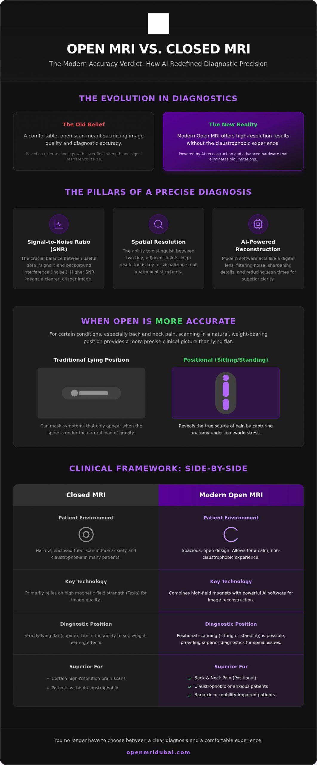
    High-quality diagnostics rely on several technical factors that go beyond the machine’s appearance. The signal-to-noise ratio (SNR) is perhaps the most critical. It represents the balance between the “signal” captured from your body’s tissues and the background electronic “noise.” A higher SNR results in a clearer, crisper image that allows for better visualization of small structures. Spatial resolution also plays a vital role, as it determines the system’s ability to distinguish between two tiny, adjacent points. Finally, the expertise of the radiologist interpreting the data is paramount. Even the highest-resolution scan requires a skilled professional to identify subtle pathologies that might indicate an underlying health issue.

    The Traditional View: Why Closed Was Considered Superior

    The belief that closed systems were inherently better stems from the physics of cylindrical magnets. These “closed-bore” machines typically offer higher Tesla (T) ratings, which measure magnetic field strength. In the past, older open magnets often struggled with “fringe” interference. This occurred because the magnetic field wasn’t as tightly contained as it is in a tube, sometimes leading to grainier images. While these historical limitations were real, they no longer apply to advanced diagnostic centers in the UAE. Today’s high-field open magnets, combined with sophisticated shielding and computational power, have effectively eliminated these old interference issues. You don’t have to choose between a clear diagnosis and a comfortable experience anymore.

    How AI and Field Strength Work Together for High-Resolution Results

    In the past, image quality was limited by the physical strength of the magnet. If you wanted high-resolution results, you generally had to enter a high-Tesla closed machine. Today, that’s no longer the case. The integration of artificial intelligence has revolutionized how we process diagnostic data. It allows us to answer the question, is open MRI as accurate as closed MRI, with a resounding yes for the vast majority of clinical applications. By shifting the focus from raw hardware power to sophisticated software reconstruction, we’ve unlocked a new era of patient-centric diagnostics.

    One major hurdle for older open machines was “artifacts,” or blurring caused by slight patient movements or lower signal levels. Modern AI-powered diagnostic imaging acts as a digital lens, filtering out this background noise and reconstructing raw data into sharp, high-definition visuals. This is especially vital for complex neurological cases where every millimeter of tissue detail counts. When every pixel is optimized by advanced algorithms, the diagnostic clarity matches or exceeds what was once only possible in a confined, cylindrical tube.

    The Role of AI in Modern Diagnostics

    Modern AI algorithms don’t just “clean up” an image; they actively fill in data gaps to create sharper edges and clearer contrast. This noise reduction technology allows for faster scans because the system can interpret signals more efficiently. For patients in Dubai, this means less time spent in the machine and a more reliable result for the radiologist. By highlighting subtle abnormalities that might have been missed by older, hardware-only systems, AI ensures that your diagnostic journey is both comfortable and precise. It’s a supportive approach that uses innovation to remove the traditional trade-off between comfort and quality.

    Field Strength vs. Image Quality

    There’s a common medical myth that “more Tesla always equals a better diagnosis.” While a 3.0T machine has its place in specific research or ultra-fine vascular studies, a 0.5T or 1.2T open MRI equipped with advanced software can easily rival the output of a standard 1.5T closed machine. AI algorithms intelligently compensate for lower field strengths by reconstructing high-definition visuals from complex signal data. This synergy between hardware and software is what defines the modern AI-powered diagnostic imaging experience. It ensures that you receive the highest standard of care without the anxiety of a confined space, proving that the smartest technology is the one that puts the patient first.

    When Open MRI Is More Accurate: The Power of Positional Scanning

    While we’ve established that AI closes the gap in image resolution, there are specific clinical scenarios where the open design provides a distinct diagnostic edge. When patients ask, is open MRI as accurate as closed MRI, they are often surprised to learn that for spinal and joint issues, the open format can actually be superior. This is due to the capability of performing a sitting open mri dubai, which allows the body to be imaged in its natural, symptomatic state. For many, pain isn’t something that happens while lying down; it’s something that occurs during the daily activities of sitting, standing, or bending.

    Standard closed machines require you to lie perfectly flat. While this is fine for many internal organ scans, it’s often counterproductive for diagnosing chronic back or neck pain. Many pathologies only manifest when the body is under the stress of gravity. By utilizing positional scanning, we can capture the true state of your anatomy while you are sitting or standing. This provides a more precise clinical picture than a supine scan ever could, ensuring that your treatment plan is based on how your body actually functions in the real world.

    The Weight-Bearing Advantage

    Gravity is a powerful diagnostic variable. When you lie down, the pressure on your spinal discs is relieved, which can cause a herniation to temporarily recede. This often leads to “false negative” results where a scan looks normal despite the patient being in significant pain. An upright scan captures the spine under the weight of the head and torso. This weight-bearing perspective is essential for identifying the true extent of disc bulges or spinal misalignments. Athletes in the UAE frequently seek out this technology because it allows for joint imaging in functional positions, revealing instabilities that stay hidden when the limb is at rest.

    Reducing Motion Artifacts Through Comfort

    The most common cause of a failed or blurry MRI isn’t the machine’s age; it’s patient movement. In a traditional, enclosed tube, even a minor bout of claustrophobia can lead to involuntary twitching or heavy breathing. These micro-movements create “motion artifacts” that degrade image quality. Because an open environment significantly reduces anxiety, patients find it much easier to remain still for the duration of the procedure. A relaxed patient in a sitting open mri dubai environment produces cleaner, more reliable data. This psychological link between physical comfort and diagnostic success is a cornerstone of modern, patient-centric care.

    Is Open MRI as Accurate as Closed MRI?

    Comparing Open vs. Closed MRI: A Clinical Framework

    Choosing between imaging methods shouldn’t be a source of stress or confusion. For approximately 95% of routine clinical diagnostics, both systems provide equivalent results that allow physicians to create effective treatment plans. If you’ve been wondering: is open MRI as accurate as closed MRI? The answer is yes for the vast majority of standard orthopedic, neurological, and abdominal scans. The decision often comes down to a balance of your personal comfort and the specific clinical questions your doctor needs to answer.

    While closed units sometimes offer slightly faster scan times due to higher raw field strength, open units provide specialized benefits that a traditional tube simply cannot match. It’s a trade-off between the pure speed of a high-Tesla machine and the specialized positioning and physical ease of an open environment. For most patients in Dubai, the reduction in anxiety and the ability to be scanned in a natural position far outweigh the few extra minutes spent in the machine.

    Best Uses for Open MRI Technology

    Open systems excel in specific scenarios where traditional machines struggle to provide a complete picture or a comfortable experience. We highly recommend this technology for several key groups:

    • Chronic Back and Neck Pain: Utilizing a sitting open mri dubai allows for weight-bearing images that reveal disc issues hidden during lying-down scans.
    • Pediatric Imaging: Children often feel frightened in enclosed spaces. The open design allows a parent to stay close and even hold their child’s hand, often removing the need for sedation.
    • Bariatric Patients: Traditional bores have strict weight and size limits. Open machines provide the necessary space for patients of all body types to be scanned with dignity and comfort.
    • Severe Claustrophobia: For those who experience intense anxiety in confined spaces, the open environment ensures the scan is completed successfully without the need for repeat appointments.

    Questions to Ask Your Radiologist

    Before you book your appointment, it’s helpful to have a direct conversation with the imaging facility. This ensures you’re receiving the highest standard of care available in the UAE. Consider asking the following questions:

    • “Does this facility utilize AI-powered diagnostic imaging to enhance the resolution of my open scan?”
    • “Can I be scanned in a sitting or standing position to better evaluate my specific pain symptoms?”
    • “What is the expected resolution for this specific body part, and will it meet my referring doctor’s requirements?”

    If you’re looking for a diagnostic experience that prioritizes both your comfort and clinical precision, you can book an open MRI scan at our Jumeirah facility to get started on your path to recovery.

    Choosing Advanced Imaging in the UAE: The Open MRI Dubai Standard

    Open MRI Dubai stands as a premier destination for AI-Powered Diagnostic Imaging in the United Arab Emirates. We’ve carefully designed our services to ensure that the question of whether an is open MRI as accurate as closed MRI is answered through innovation rather than compromise. By adhering to the latest Dubai Health Authority standards for diagnostic imaging, which were updated to ensure peak patient safety in late 2025, we provide a level of care that prioritizes both precision and comfort. You don’t have to choose between a supportive environment and the high-resolution results your physician requires for an effective treatment plan.

    We believe that modern healthcare should be as stress-free as possible. Our facility isn’t just about the machines; it’s about a philosophy of care that puts your well-being at the center of every scan. By combining the latest technological advancements with a premium, patient-centric environment, we’ve set a new standard for diagnostics in Jumeirah. This approach ensures that every individual who walks through our doors feels heard, supported, and confident in the accuracy of their results.

    The AI-Powered Difference

    Our facility integrates sophisticated AI algorithms that act as a bridge between patient comfort and clinical excellence. These tools allow our team to reconstruct raw signal data into sharp, high-definition visuals that rival traditional closed systems. This focus on diagnostic integrity ensures that every patient, regardless of their condition, receives a scan that is both precise and reliable. We believe that a premium experience should include the peace of mind that comes from knowing your data is being processed by some of the most advanced technology in the region. Our team acts as a technological caregiver, guiding you through the process with a focus on your physical and emotional comfort.

    Booking Your Comfortable Scan

    Moving from a referral to a completed scan is straightforward and stress-free. When you arrive for your sitting open mri dubai session in Jumeirah, you’ll find an environment that feels more like a modern suite than a cold, traditional clinic. Our technicians are trained to provide empathetic support, ensuring you feel well-informed throughout the procedural journey. Whether you’re seeking a scan for a complex spinal pathology or a routine orthopedic check, our open-bore and sitting units offer the space you need to remain calm. This methodical approach ensures that we capture the highest quality data while you remain in a natural, relaxed position.

    Experience high-resolution, non-claustrophobic imaging at Open MRI Dubai.

    A Clear Path to Diagnostic Clarity and Comfort

    The evolution of medical imaging has reached a point where you no longer have to sacrifice peace of mind for clinical precision. We’ve explored how AI-powered technology and specialized sitting MRI systems have fundamentally changed the answer to the question: is open MRI as accurate as closed MRI? By combining advanced software with positional scanning, modern open systems provide the high-resolution data your doctor needs while keeping you in a relaxed, non-claustrophobic environment. This synergy ensures that weight-bearing issues are captured with absolute clarity. These vital details are often hidden in traditional, lying-flat machines.

    Our facility in Jumeirah is dedicated to this standard of excellence. With expert radiologists specializing in open-format diagnostics and a focus on patient-centric care, we ensure your diagnostic journey is as seamless as it is accurate. You deserve a healthcare experience that respects your physical comfort without compromising on the quality of your results. Take the next step toward understanding your health in a space designed for your well-being. Book your high-resolution Open MRI scan today and discover how our specialized technology provides the answers you need with the comfort you deserve.

    Frequently Asked Questions

    Is the image quality of an open MRI good enough for a spine surgery referral?

    Yes, the image quality provided by our advanced open systems is more than sufficient for spine surgery referrals. Surgeons in Dubai frequently request these scans because they can evaluate the spinal column under natural, weight-bearing conditions. This provides a more precise assessment of disc herniations or spinal stenosis that might temporarily recede when a patient is lying flat in a traditional machine, ensuring a more accurate surgical plan.

    How does AI help in making an open MRI more accurate?

    AI technology enhances accuracy by processing raw data to reduce background noise and sharpen image edges. These advanced algorithms help bridge the gap when patients ask if an is open MRI as accurate as closed MRI by reconstructing high-definition visuals from complex signals. The software compensates for lower hardware field strengths, ensuring that the final diagnostic report is clear and reliable for your referring physician.

    Can I get a sitting MRI if I have severe claustrophobia?

    A sitting MRI is an ideal solution for patients with severe claustrophobia. Unlike traditional machines that require you to enter a narrow, enclosed tube, our open design allows you to remain in a comfortable seated position with a clear view of your surroundings. This significantly reduces anxiety and the feeling of confinement, which often eliminates the need for sedation or the risk of repeat appointments due to patient movement.

    Do doctors in the UAE accept results from an open MRI scan?

    Doctors across the UAE widely accept and trust results from high-quality open MRI scans. Our facility adheres to the strict diagnostic imaging standards set by the Dubai Health Authority (DHA), which were updated in late 2025 to ensure peak patient safety. Whether you are seeing a specialist in Jumeirah or a surgeon at a major hospital, these reports provide the authoritative data needed for your healthcare journey.

    What is the difference in Tesla strength between open and closed MRI?

    Traditional closed machines typically operate at field strengths of 1.5T to 3.0T, while modern open systems generally range from 0.5T to 1.2T. A common question is whether an is open MRI as accurate as closed MRI despite this difference. The integration of AI-powered imaging allows modern open units to achieve diagnostic results that rival their higher-Tesla counterparts, ensuring clinical precision even if the raw magnetic field strength is lower.

    Is an open MRI as fast as a traditional closed MRI scan?

    Modern open MRI scans are comparable in speed to traditional sessions, typically lasting between 15 and 60 minutes depending on the area being examined. While some high-field closed machines might offer slightly shorter scan times for specific sequences, the difference is usually negligible for the patient. The added comfort of an open environment often makes the time feel like it passes much faster than in a confined tube.

    Are there any body parts that cannot be scanned with an open MRI?

    Most body parts, including the spine, joints, brain, and abdomen, can be effectively scanned using an open MRI system. There are very few clinical exceptions where a high-field closed bore is mandatory, such as certain ultra-fine vascular studies or specific research-level cardiac imaging. For the vast majority of orthopedic and neurological diagnostic needs in Dubai, an open system is a perfectly valid and highly accurate choice.

    Does a sitting MRI show things a lying-down MRI might miss?

    Yes, a sitting MRI can reveal pathologies that a standard lying-down scan might miss. When you lie flat, gravity is removed from your spine and joints, which can cause disc bulges to recede or joint instabilities to disappear. Capturing images while you are in a seated, weight-bearing position provides a more realistic view of how your anatomy reacts to the daily stress and pain triggers you experience.

  • Open MRI Dubai: The Complete Guide to AI-Powered Imaging and Patient Comfort

    Open MRI Dubai: The Complete Guide to AI-Powered Imaging and Patient Comfort

    What if you could receive a crystal-clear diagnostic image without ever stepping foot inside a narrow, enclosed tunnel? For many patients across the UAE, the prospect of a traditional scan triggers immediate anxiety. You might worry about the feeling of confinement, or perhaps you find it physically impossible to lie flat for long periods due to chronic pain. It’s a common concern that often leads to delayed diagnoses or the unnecessary stress of sedation.

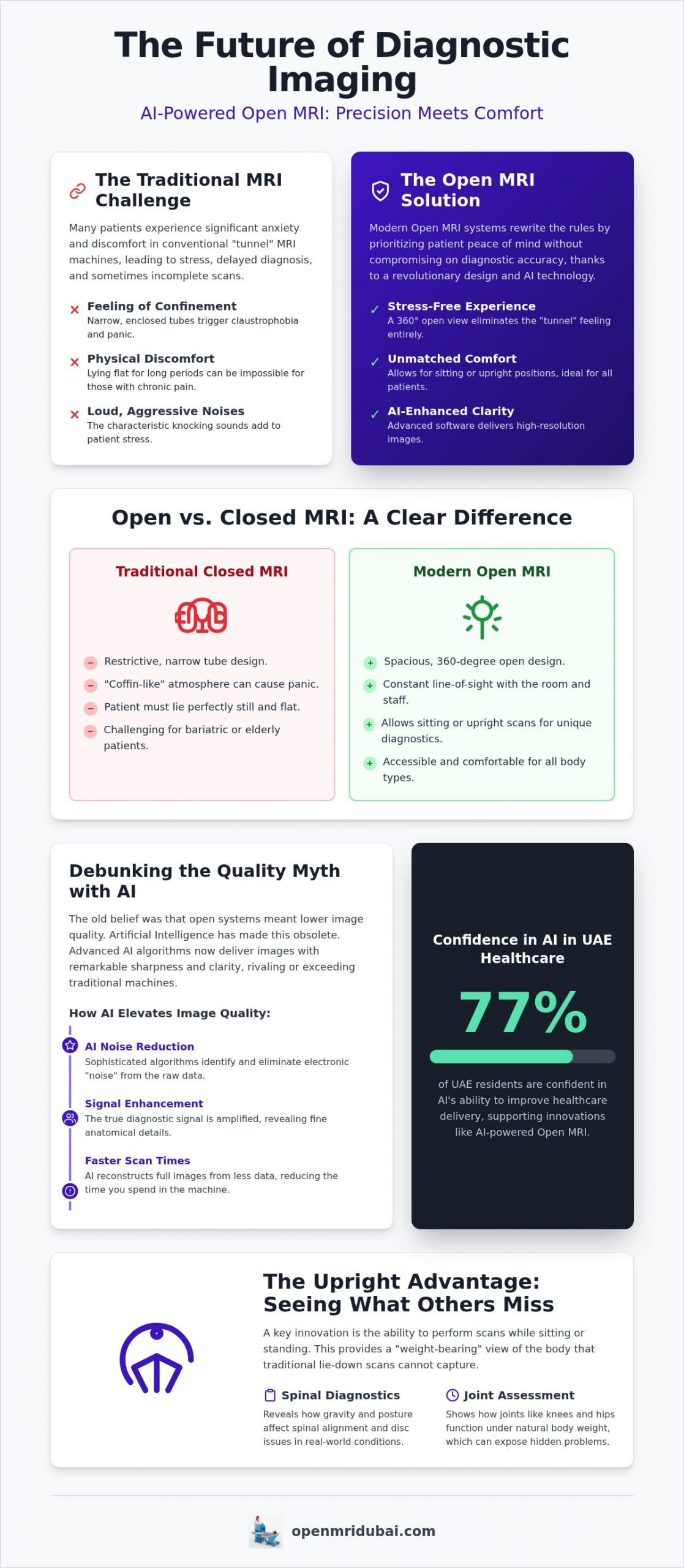
    We understand that your peace of mind is just as important as the accuracy of your results. This guide explores how the latest advancements in Open MRI Dubai technology are rewriting the rules of diagnostic care by combining AI-enhanced precision with the physical freedom of sitting or upright positioning. Recent data indicates that 77% of UAE residents are confident in AI’s ability to improve healthcare delivery, and this innovation is now making the diagnostic journey more comfortable than ever. You’ll learn how these sophisticated systems deliver high-resolution clarity that rivals traditional machines while ensuring your comfort remains the absolute priority. We will walk you through the benefits of AI-powered imaging and explain why a patient-centric approach means you no longer have to sacrifice clinical quality for a stress-free experience.

    Key Takeaways

    • Understand how the spacious, 360-degree design of an open MRI Dubai scan eliminates the restrictive “tunnel” feeling while maintaining strict clinical standards.
    • Discover how AI-powered signal enhancement ensures high-resolution diagnostic clarity, proving that patient comfort doesn’t have to come at the expense of image quality.
    • Learn about the unique benefits of sitting or upright MRI technology for capturing weight-bearing details in the spine and joints that traditional scans might miss.
    • Prepare effectively for your visit by understanding UAE safety protocols and the simple steps you can take to ensure a smooth, stress-free imaging session.
    • Gain insights into choosing a diagnostic partner that prioritizes both technological innovation and an empathetic environment for a truly premium experience.

    What is an Open MRI Dubai Scan? Defining Modern Diagnostic Comfort

    Modern medical imaging has undergone a significant transformation. An open MRI Dubai scan represents a departure from the traditional, restrictive “tunnel” experience. Unlike standard systems that require you to enter a narrow tube, an open system uses a design featuring two parallel plates or a C-arm structure. This configuration provides a 360-degree view of the room, ensuring you never feel trapped or isolated. It’s an ideal solution for those who prioritize their mental well-being alongside their physical health. The evolution of this technology has moved beyond simple “wide-bore” tunnels, which are essentially just slightly larger tubes. Today, truly open configurations allow for sitting or upright positions, offering a level of freedom that was previously impossible in clinical settings.

    Understanding the physics of Magnetic Resonance Imaging (MRI) helps clarify why this design is so revolutionary. Traditional machines rely on a closed-bore magnet to create the necessary field. However, modern engineering allows us to achieve high-quality results using open-format hardware. This shift is vital for reducing the physiological stress response. Patients often find that the open environment eliminates the need for pharmaceutical assistance to manage anxiety. When you’re relaxed, your heart rate remains steady and involuntary movements decrease. This cooperation between patient and technology results in diagnostic excellence without the typical hurdles of traditional radiology.

    The Core Differences: Open vs. Closed Bore Systems

    The most striking difference is the spatial environment. Closed-bore systems are often described as having a “coffin-like” atmosphere, which can be distressing. In contrast, the open format eliminates this feeling of confinement. This accessibility is vital for bariatric patients who may not fit comfortably in a standard tube, as well as elderly patients who require more physical room to feel secure. Open systems are also significantly quieter, removing the aggressive knocking sounds that characterize older technology.

    Addressing the Claustrophobia Factor

    For many, a trip to the radiologist is overshadowed by the fear of enclosed spaces. An open MRI Dubai facility addresses this “closed-in” panic trigger by maintaining a permanent line-of-sight with the outside world. You can see the technician or even a family member throughout the procedure. This visual connection drastically increases success rates for patients who previously failed to complete scans in closed machines. It’s a supportive approach that transforms a stressful medical requirement into a manageable, calm experience.

    AI-Powered High-Resolution Imaging: Debunking the Quality Myth

    Traditional views on diagnostic imaging often suggest a trade-off exists between patient comfort and image clarity. For years, the assumption was that an open MRI Dubai facility could not match the raw power of a closed-bore system. Artificial Intelligence has fundamentally dismantled this myth. In the past, open magnets relied solely on their hardware’s field strength, which could sometimes result in “grainy” images. Today, advanced AI algorithms act as a sophisticated digital lens. These systems identify and remove electronic “noise” in real-time, while simultaneously enhancing the diagnostic signal to produce images of remarkable sharpness.

    This computational power allows for significantly faster acquisition. Patients now spend less time in the scanning area because the AI can reconstruct full, detailed images from less raw data. This efficiency doesn’t just improve your experience; it ensures that even the slightest involuntary movement doesn’t compromise the scan quality. It’s a shift from hardware-heavy solutions to software-driven precision, ensuring every minute spent during your session translates into actionable medical data. If you’ve ever wondered is open MRI as accurate as closed MRI, the answer lies in how these AI-driven advancements have effectively closed the gap between the two technologies.

    Signal-to-Noise Ratio (SNR) and AI Reconstruction

    The technical secret to high-resolution results lies in the Signal-to-Noise Ratio. AI reconstruction software “cleans” the magnetic data, separating essential anatomical information from background interference. We’ve moved into an era where software-driven precision compensates for the physical design of the magnet. When comparing an AI-enhanced open scan to a standard closed MRI, the clarity is often indistinguishable to the trained eye. This ensures that your open MRI scan provides the exact level of detail your physician requires for an accurate assessment.

    Diagnostic Confidence for Specialists

    Specialists in neurology and orthopaedics now rely on these enhanced scans for critical decision-making. AI helps highlight micro-lesions, subtle ligament tears, and minute tissue changes that are vital for accurate surgical planning. The precision offered by these systems gives doctors the confidence to diagnose complex conditions without hesitation. AI-powered imaging serves as the essential bridge between total patient comfort and uncompromising diagnostic excellence. By utilizing these tools, clinicians can identify issues that might have been overlooked in the past, ensuring that your treatment plan is based on the most accurate visual evidence available in modern medicine.

    Sitting and Upright MRI: Transforming the Diagnostic Experience

    Pain is often positional. If your back hurts specifically when you sit or stand, a scan taken while you’re lying flat might not tell the whole story. This is where sitting MRI technology changes the diagnostic landscape. Unlike traditional machines that require a recumbent position, these systems allow you to be scanned in a natural, upright posture. It’s a significant breakthrough for orthopaedic diagnostics because it captures the body exactly as it functions in daily life, under the natural pressure of gravity.

    The experience is remarkably different from a standard clinical encounter. You aren’t slid into a dark tube; instead, you sit comfortably in a padded chair. Many patients find they can even watch television or remain in a natural posture that doesn’t aggravate their symptoms. This relaxed environment doesn’t just reduce anxiety; it ensures you stay still, which leads to superior image quality. For those seeking a premium open MRI Dubai solution, this upright capability represents the pinnacle of patient-focused design.

    Who Benefits from Sitting MRI Scans?

    Certain conditions make traditional scanning nearly impossible for some individuals. A seated position offers a dignified and comfortable alternative for several groups:

    • Patients with severe sciatica or lumbar stenosis often find lying horizontally to be physically unbearable, as the flat position can intensify nerve pain.
    • Individuals with respiratory issues or heart conditions often breathe more easily when seated upright, making the duration of the scan much safer and more comfortable.
    • Children feel much more secure when they can sit in a familiar posture and maintain eye contact with their parents, which significantly reduces the need for medical sedation.

    Weight-Bearing vs. Recumbent Imaging

    There’s a critical clinical difference between being “unloaded” and “loaded.” When you lie down for a traditional scan, gravity’s pressure on your spine and joints is removed. This can cause a disc herniation to temporarily retract or “disappear” on the image, leading to an incomplete diagnosis. Upright imaging applies the “Gravity Factor,” revealing the true extent of nerve compression or spinal instability that occurs when you are standing or walking.

    This approach is also highly effective for assessing knee and hip joints. By scanning the joint while it’s supporting your body weight, radiologists can see how the cartilage and ligaments actually behave under stress. It provides a level of detail that recumbent imaging simply cannot match. For anyone exploring their options for an open MRI Dubai, choosing a weight-bearing scan can be the decisive factor in creating an accurate and effective long-term recovery plan.

    Open MRI Dubai: The Complete Guide to AI-Powered Imaging and Patient Comfort

    Preparing for Your Scan: Safety Protocols and UAE Standards

    Your safety is the foundation of every procedure we perform. While the design of an open MRI Dubai system is physically welcoming, it still utilizes a powerful magnetic field to generate detailed images. This means that preparation is just as rigorous as it would be for a traditional scan. You’ll be asked to remove all metallic objects, including jewelry, watches, and hairpins. We recommend wearing comfortable, loose-fitting clothing without zippers or metal buttons. If your outfit isn’t suitable, we’ll provide a high-quality gown to ensure your comfort and the integrity of the scan results.

    Medication and diet usually don’t need to change unless your physician specifies otherwise. If your scan requires a contrast agent to highlight specific tissues, you might be asked to fast for a few hours beforehand. Your radiographer will guide you through every step, acting as your professional partner to ensure a stress-free experience. They are trained to monitor you throughout the session, providing reassurance and answering any questions you have about the technology or the process.

    The Screening Process

    Before you enter the imaging suite, you’ll complete a comprehensive health questionnaire. This step is vital for identifying internal medical devices like pacemakers, cochlear implants, or certain types of vascular clips. Keep in mind that ‘open’ refers to the machine’s architecture, not a lack of magnetic force. For children or elderly patients, we take extra time to explain the rhythmic sounds of the hardware. Understanding what to expect helps reduce the startle response and ensures a calm environment for everyone involved.

    National Healthcare Quality Standards

    Quality in the UAE is governed by strict regulatory frameworks. Our facility adheres to the latest Dubai Health Authority (DHA) standards for diagnostic imaging, which were updated on November 26, 2025. These regulations ensure that every piece of equipment is calibrated for precision and that patient safety protocols are followed without compromise. Choosing a DHA or MOHAP licensed facility means your results are analyzed by verified experts using the highest clinical benchmarks. This commitment to excellence ensures your reporting is accurate and delivered within professional timelines, giving you and your doctor the clarity needed for your care plan. If you’re ready to experience this level of professional care, you can schedule your open MRI Dubai session today.

    Selecting an Imaging Partner: Why Technology and Empathy Matter

    Choosing the right diagnostic partner is a decision that impacts both your mental comfort and your physical recovery. While many facilities offer imaging, a specialized center for an open MRI Dubai provides a unique environment tailored specifically to those who find traditional machines challenging. In a hospital, MRI suites are often high-traffic areas dealing with urgent emergencies, which can feel cold and rushed. A dedicated open-imaging center prioritizes a calmer pace. This specialization ensures that every member of the staff, from the front desk to the radiographer, understands how to support patients with claustrophobia or chronic pain.

    Clinical professionalism shouldn’t feel detached. By combining premium facility design with advanced computational tools, we create a space where you feel like a guest rather than a patient. This “Technological Caregiver” approach means your well-being is the primary metric of success. The journey doesn’t end when the scan is finished. The integration of AI doesn’t just improve image quality; it also streamlines the reporting process. AI-assisted analysis helps radiologists identify key findings faster, ensuring your specialist receives a comprehensive report without unnecessary delays. This efficiency is a core part of our commitment to your health journey.

    The Technological Caregiver Approach

    A superior diagnostic experience balances high-end AI tools with a supportive atmosphere. Specialized centers often feel more like a wellness environment than a traditional clinic. This relaxed setting is particularly beneficial for those who’ve had negative experiences with medical diagnostics in the past. We believe in transparent communication, explaining every detail of the procedure so you never feel uncertain. This clarity builds trust and ensures that you remain relaxed throughout your open MRI Dubai session. By focusing on the patient’s emotional state, we achieve better cooperation and clearer results.

    Next Steps: Booking Your AI-Powered Scan

    If you’re ready to move forward, the process is straightforward and efficient. You can obtain a referral from your primary physician or book a private consultation directly with our team. While we specialize in open MRI technology, we also offer a full suite of diagnostic tools, including ct scan dubai and xray dubai services. This allows for a comprehensive diagnostic profile within a single, comfortable location. Our team is dedicated to providing a seamless journey from your initial booking to the final delivery of your results. Don’t let anxiety delay your diagnosis any longer. Schedule your comfortable Open MRI scan today and discover the difference that empathetic, technology-driven care can make.

    Empower Your Health with Precision and Peace of Mind

    Diagnostic clarity shouldn’t come at the cost of your mental well-being. By choosing an open MRI Dubai experience, you’re opting for a methodology that respects your comfort without compromising on clinical precision. We’ve explored how advanced AI-powered imaging reconstruction now delivers high-resolution results that rival any traditional system. Specialized sitting and upright MRI technology further ensures that positional pain is accurately captured under real-world weight-bearing conditions. These innovations, paired with the expertise of our nationally accredited radiologists, mean you can approach your diagnosis with total confidence.

    Our facility is designed to be a sanctuary for those who prioritize a supportive, empathetic environment. We adhere strictly to UAE healthcare regulations to ensure your safety remains paramount at every stage. You don’t have to let the fear of confinement or the worry of an incomplete scan hold you back from the answers you deserve. Take the next step toward a clearer, more comfortable healthcare journey today.

    Book your AI-powered Open MRI at our specialized centre and experience a new standard of diagnostic excellence. Your path to recovery starts with a stress-free experience.

    Frequently Asked Questions

    Is an open MRI as accurate as a traditional closed MRI?

    Modern open systems provide high-resolution results that match traditional machines through advanced computational power. By using AI-powered diagnostic imaging, the software removes electronic noise and reconstructs data for maximum clarity. This ensures your specialist receives precise information for neurology or orthopaedic assessments. The gap in quality that once existed between open and closed systems has been effectively bridged by these sophisticated digital tools, ensuring your diagnosis is both accurate and reliable. For a deeper clinical breakdown, explore our detailed guide on whether open MRI is as accurate as closed MRI to understand exactly how modern technology has transformed diagnostic precision.

    Can I sit or stand during my MRI scan in Dubai?

    You can absolutely choose a sitting position for your scan at specialized facilities in the city. This technology allows for upright diagnostic imaging, which is particularly useful for identifying spinal issues that might be missed while lying flat. While standing options are available in some configurations, sitting is the most common upright choice. It provides a natural posture that’s more comfortable for those with chronic back pain, joint instability, or respiratory difficulties.

    Do I need sedation for an open MRI if I am claustrophobic?

    Most patients find they don’t need sedation when using an open MRI Dubai system. The 360-degree view and lack of a restrictive tunnel significantly reduce the physiological triggers for anxiety. You can maintain a constant line of sight with your radiographer and even watch television during the procedure. This supportive environment helps you remain calm and still, which is essential for capturing high-quality diagnostic images without the side effects of sedative medication.

    How long does a typical open MRI scan take with AI technology?

    A typical scan usually lasts between 15 and 45 minutes, depending on the specific area being examined. AI technology plays a crucial role in reducing this time by allowing the machine to acquire data more efficiently. Because the software can reconstruct detailed images from less raw information, you spend less time in the scanning area. This faster acquisition doesn’t compromise detail; it simply makes the entire process more streamlined and convenient for your busy schedule.

    Are there weight limits for the sitting open MRI machine?

    Open and sitting machines generally accommodate higher weight limits than traditional closed-bore systems. While specific limits can vary by machine model, many systems are designed to support patients up to approximately 225 kilograms. The spacious, open design also provides much more physical room for bariatric patients who might find a standard tube too restrictive. It’s best to confirm the exact capacity with the facility when booking your appointment to ensure the equipment meets your needs.

    Will my health insurance cover an open MRI scan?

    Most major health insurance providers in the UAE cover open MRI scans when they’re medically necessary. Since health insurance is mandatory for all Dubai residents under Law No. 11 of 2013, you should check your specific policy for co-payment details. Some plans might require prior authorization from your referring physician. Our administrative team can help verify your coverage and explain any applicable prescription co-pay caps that might affect your diagnostic visit.

    Can I have an open MRI if I have metal implants or a pacemaker?

    Safety depends entirely on the specific type and material of your implant. While some modern pacemakers and implants are “MRI-conditional,” others are strictly incompatible with magnetic fields. You’ll need to provide the specific make and model of your device during the screening process. Our radiographers follow strict DHA safety protocols to evaluate every case individually. If an MRI isn’t safe for you, we can discuss alternative diagnostic options like a CT scan.

    What is the main difference between open MRI and wide-bore MRI?

    The main difference lies in the physical structure and the patient’s field of vision. A wide-bore MRI is still a tunnel, though it has a slightly larger diameter of 70 centimeters compared to the standard 60 centimeters. A true open MRI Dubai scan uses a C-arm or parallel plate design that’s completely open on the sides. This provides a full 360-degree view, which is much more effective for patients with severe claustrophobia who cannot tolerate any enclosure.