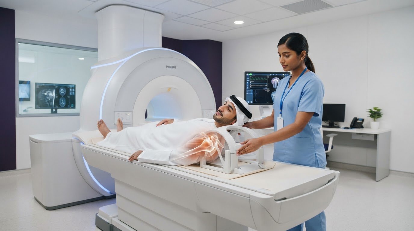
Could the source of shoulder pain be in structures that an X-ray cannot show clearly? When symptoms persist or an injury needs closer assessment, a clinician may recommend a comprehensive shoulder MRI scan to examine soft tissues and other structures in detail. You may be wondering why MRI is the next step, what it could reveal, or whether staying still in the scanner will feel uncomfortable.
This guide explains why a clinician may recommend a shoulder MRI and what the images can help assess, including tendons, ligaments, cartilage and surrounding tissues. You’ll also learn what to share before the scan, how to prepare and what happens during imaging. If enclosed spaces make you uneasy, a specialized scan at Open MRI Dubai may offer a more comfortable environment. Your referral and the clinical question guide the imaging plan.
After the scan, a radiologist interprets the images and prepares a report for your referring clinician. Your clinician discusses the findings alongside your symptoms and examination. Knowing how these steps fit together can help you feel more prepared for your appointment.
Key Takeaways
- A clinician recommends a shoulder MRI based on your symptoms, examination, medical history and the question the scan needs to answer.
- MRI uses magnetic fields and radiofrequency signals to create views of structures inside and around the shoulder joint.
- Open-format scanners have a different physical design that may feel more comfortable for some people. The imaging plan still depends on the clinical need.
- Share relevant medical history, implant information, previous imaging and referral details with the imaging team, and follow their preparation instructions.
- If you’re considering a shoulder mri dubai scan, remember that the radiologist’s report and your clinician’s assessment work together to guide next steps.
Why might a clinician recommend a shoulder MRI scan?
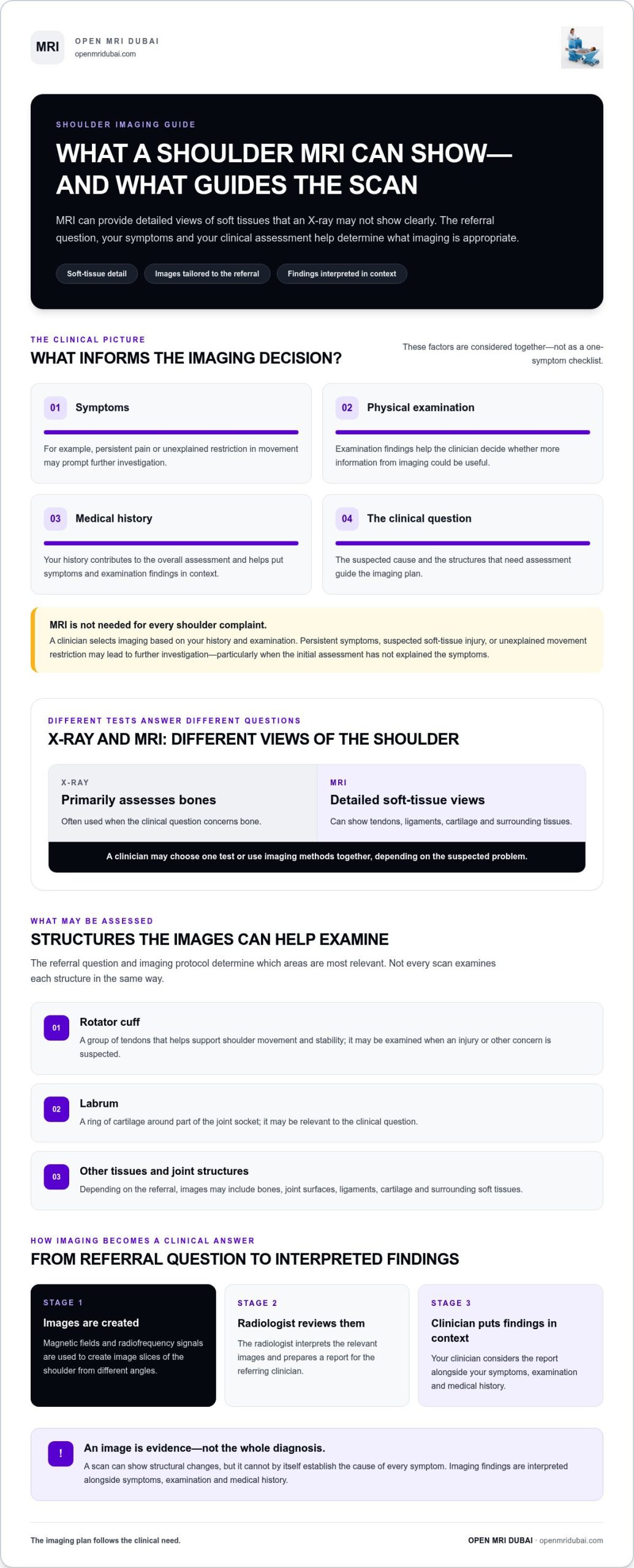
A shoulder MRI, or magnetic resonance imaging scan, uses magnetic fields and radiofrequency signals to create detailed images of structures inside and around the joint. An X-ray is primarily used to assess bones, while MRI can provide views of soft tissues such as tendons, ligaments and cartilage. A clinician may recommend MRI when images of those structures could help answer a specific question about your shoulder.
The decision depends on the full clinical picture: your symptoms, physical examination, medical history and the suspected cause of the problem. Persistent pain, a suspected soft-tissue injury or unexplained restriction in movement may prompt further investigation, particularly if the initial assessment hasn’t explained your symptoms. Not every shoulder complaint requires MRI. The referring clinician selects imaging based on what they need to assess.
For people researching shoulder mri dubai, understanding the purpose of the scan can make a referral feel less uncertain. MRI is one part of an investigation, not a replacement for a consultation or examination.
What questions can a shoulder MRI help investigate?
Depending on the clinical question and imaging protocol, a radiologist may assess the shoulder’s tendons, ligaments, cartilage and other soft tissues. The rotator cuff, a group of tendons that helps support shoulder movement, may be examined when an injury or another concern is suspected. The labrum, a ring of cartilage around part of the joint, may also be relevant.
For example, a clinician might request MRI to investigate ongoing pain or reduced movement when they need more information about soft tissues than an X-ray can provide. Conditions such as shoulder impingement syndrome can be associated with shoulder pain, but symptoms and imaging findings need careful interpretation. A scan can show structural changes; it cannot, by itself, establish the cause of every symptom.
When might MRI be considered alongside other imaging?
Different imaging tests answer different questions. X-rays are commonly used to assess bones, while MRI can offer detailed views of soft tissues. Depending on the suspected problem, a clinician may choose one test or use imaging methods together. The appropriate choice depends on your history and examination, not on a single symptom or a general preference for one scan type.
An imaging test provides evidence about structures. A complete clinical diagnosis brings that evidence together with symptoms, examination and medical history. A finding on an image may not fully explain how your shoulder feels or moves. Your referring clinician interprets the radiology report in context and explains what the findings mean for your care.
How does a shoulder MRI create detailed images of the joint?
An MRI scanner uses a strong magnetic field and radiofrequency signals to create images of the body. In simple terms, these signals interact with tissues in different ways, and the scanner records the responses. A computer processes that information into image slices, giving the radiologist views through the shoulder from different angles.
These views let the radiologist examine the area relevant to the referral rather than relying on a single flat picture. The shoulder includes the glenohumeral joint, where the rounded top of the humerus, or upper arm bone, meets the socket of the shoulder blade. The scan can show this joint and nearby tissues. The precise images and sequences are selected according to the referral question and clinical judgment.
Which shoulder structures may appear in the images?
The scan may include bones, joint surfaces and surrounding soft tissues. The rotator cuff is a group of tendons that supports shoulder movement and stability. The labrum is a ring of cartilage around the joint socket. It may be assessed when relevant to the clinical question, but not every scan examines each structure in the same way. The radiologist reviews the images in light of the requested assessment.
If you’re learning about shoulder mri dubai, it helps to know that the scan isn’t simply a general picture of the whole shoulder. Its image views are planned to address the reason for imaging. For a patient-friendly overview of the scan and its role, see UCSF Health’s Shoulder MRI Scan: What It Shows and What to Expect.
What does AI-assisted imaging mean for patients?
Open MRI Dubai provides AI-powered diagnostic imaging, including open MRI scans. AI-powered imaging is part of the imaging service, while the images still require professional review. It does not replace a radiologist’s interpretation or a clinician’s assessment of what the findings mean for an individual patient.
Imaging technology helps produce images; clinical interpretation puts those images in context. The radiologist reviews the relevant views and prepares a report. The referring clinician relates the findings to your symptoms and examination. Professional interpretation remains central to understanding the images.
Knowing that image views are tailored to the clinical question can make the process feel more purposeful. If you’re exploring an open MRI scan, learning about the open-format approach can help you understand the imaging environment as well as the technology used to create the images.
Open-format shoulder MRI: comfort benefits and practical considerations
An open-format MRI has a more open physical design than an enclosed scanner, which may feel less confining for some patients. This can be reassuring if enclosed spaces make you anxious or you simply prefer a more open environment. Comfort is personal, however. The scanner’s design is only one part of the experience and cannot guarantee that every person will feel relaxed during an examination.
Open MRI Dubai provides open-format imaging, including sitting open MRI technology. The setup for a shoulder examination depends on the clinical question and imaging protocol. A scanner’s appearance alone doesn’t establish whether it can meet the needs of a particular examination or whether it is suitable for a specific patient.
What can an open MRI environment feel like?
Rather than being surrounded by a narrow tunnel, patients may have a more open space around them. That difference can make the scan feel more manageable for some people who dislike enclosed areas. The shoulder still needs to be positioned for the requested images, and you’ll need to follow the imaging team’s instructions and remain as still as you comfortably can while images are captured.
People respond differently to the scanner environment, positioning and sounds during imaging. If you feel uneasy, tell the imaging team so they can explain what to expect and guide you through the process. An open design may support comfort, but it doesn’t remove the need for careful positioning or make the experience identical for every patient.
How should patients think about scan suitability?
Suitability depends on the body area being examined, the clinical question and the protocol selected by the imaging team. For a shoulder MRI, the requested assessment helps determine which images are needed. The scanner format alone doesn’t establish whether the examination will answer that question. Follow the instructions for your scan and share any relevant concerns or information that may affect positioning.
If you’re comparing options for shoulder mri dubai, consider more than whether a scanner looks open or enclosed. The purpose of the examination and its imaging requirements matter just as much as the physical environment. A format that feels appealing may not suit every examination, and open MRI shouldn’t be assumed to produce the same images for every clinical need.
For a broader look at the open-format experience and comfort considerations, explore this complete guide to open MRI imaging and patient comfort. It explains the environment; your referral and imaging instructions remain the guide for your individual examination.
How to prepare for a shoulder MRI scan and what happens during it
A little preparation can make your appointment more straightforward. Follow the imaging centre’s specific instructions, as preparation and scan protocols can vary. Share relevant health information before the scan, especially details about implants or previous procedures. This helps the imaging team complete safety screening and plan the examination.
Use this sequence as a guide, while treating the instructions for your own examination as the priority:
- Gather your documents. Bring your referral information and any previous shoulder images or reports that are available and relevant.
- Tell the team about your health history. Mention implants, medical devices, past procedures and health conditions that may affect your scan.
- Choose suitable clothing. Follow the centre’s directions about clothing and metal. You may be asked to change and remove metallic items, such as jewellery or a watch, before imaging.
- Ask about anything unclear. Raise questions about preparation, positioning or what to expect with the imaging team before the scan begins.
What should you tell the imaging team beforehand?
Give specific information about any implant or device, and share relevant details about previous procedures or medical conditions. If you have earlier shoulder imaging, bring it when available so the team has useful context. Your referral information also helps identify the clinical question behind the examination. Metal screening is a routine MRI safety step; accurate information helps staff plan the imaging appropriately.
What happens while the images are taken?
The imaging team will guide you into a position for the shoulder examination and explain how to stay still while images are captured. Careful positioning helps produce useful images. The scan may include multiple image sequences, with the protocol shaped by the referral question and clinical judgment. The team will guide you through the process, so you don’t need to know the sequence in advance.
Staying still can be challenging if your shoulder is uncomfortable. Tell the staff if you’re struggling to maintain the position or feel uneasy. They can guide you and explain what they need you to do. Avoid changing position without instructions, as movement may affect the images and a sequence may need to be repeated.
If you’re preparing for a shoulder mri dubai appointment, following your specific instructions and sharing relevant information can help the team plan the examination. Explore open MRI imaging to learn about the centre’s open-format approach.
What happens after your shoulder MRI at Open MRI Dubai?
After the images have been captured, a radiologist reviews them and prepares a report for the clinician who referred you. The report describes relevant imaging observations; it isn’t, by itself, a complete explanation of your symptoms or a treatment plan. Your referring clinician brings the imaging information together with your medical history and examination.
Open MRI Dubai provides open-format imaging and AI-powered diagnostic imaging. These are part of the imaging service, while the radiologist’s interpretation and your clinician’s assessment remain central to understanding what the findings mean for you.
Who explains the shoulder MRI findings?
The radiologist interprets the images and writes the report. Your treating clinician discusses the findings with you and considers them alongside your symptoms, physical examination and health history. An image may show a structural feature, but its significance depends on the overall clinical picture. Your clinician can explain how unfamiliar terms in the report relate, or may not relate, to what you’re experiencing.
The scan supplies information, and your clinician uses it as part of your care discussion. Avoid drawing conclusions or making treatment decisions from images alone. If you have questions about the report, raise them with the clinician managing your shoulder concern, who can interpret the findings in context.
How can you take the next step?
If you have a referral, use it to arrange the imaging your clinician has indicated. The referral helps identify the purpose of the examination, and the imaging team uses that information to support the scan process. Once the report is available to your referring clinician, follow up with them to discuss the findings and any appropriate next steps.
An open-format imaging environment may feel more comfortable for some people, particularly if enclosed spaces cause unease. Comfort is individual, and scanner suitability depends on the clinical question and imaging protocol. Open MRI Dubai’s open-format approach is designed with patient comfort in mind, and AI-powered imaging is part of its imaging service. Neither the scanner format nor the technology alone determines what a scan will show or what care is right for you.
Knowing who reviews your images and how the report fits into your care can make the process clearer. If you have a referral and would like to learn about the centre’s open-format approach, explore open MRI imaging.
Take your next step with confidence
A referral starts a focused conversation about what your clinician needs to understand. Before arranging a shoulder mri dubai scan, keep your referral details and relevant medical information ready, and note any questions you’d like answered. This can help you feel prepared and understand the purpose of the examination.
If enclosed spaces make you uneasy, an open-format imaging environment may feel more comfortable, although the right approach still depends on the clinical question and scan protocol. Open MRI Dubai provides open-format imaging, including sitting open MRI technology, and AI-powered diagnostic imaging. These features are part of the imaging service; your radiologist and referring clinician remain essential to interpreting findings in context.
To learn more about Open MRI Dubai’s open-format imaging, explore open MRI imaging. If you have a referral, follow your clinician’s guidance for your examination.
Frequently Asked Questions
What can a shoulder MRI show?
A shoulder MRI can show bones, joint surfaces and soft tissues, including tendons, ligaments and cartilage. The areas examined depend on the clinical question, so the images may focus on specific structures rather than assess every part equally. A radiology report describes what is visible, but a finding doesn’t automatically explain pain or require treatment. Your clinician interprets the report alongside your symptoms and examination.
Does a shoulder MRI show rotator cuff injuries?
A shoulder MRI can provide images of the rotator cuff tendons and may help assess suspected injury. The scan’s usefulness depends on the examination protocol and the concern described in the referral. If you have pain or weakness during a particular movement, tell your clinician so they can interpret the images in context. Discuss the report with your treating clinician rather than using it alone to decide on care.
Is an open MRI suitable for a shoulder scan?
An open MRI may be suitable for some shoulder examinations, and its more open environment may feel preferable if enclosed spaces make you anxious. Suitability depends on the clinical question and imaging protocol, not simply on the scanner’s appearance. If your clinician has recommended imaging, follow the referral instructions for that examination. Open MRI Dubai provides open-format imaging, though no scanner format is right for every patient or scan.
How should I prepare for a shoulder MRI?
Follow the imaging centre’s directions and share information about implants, medical devices, past procedures or relevant health conditions before the appointment. Wear clothing that follows the centre’s guidance, and be prepared to remove metal items if asked. Bring your referral information and any earlier shoulder images or reports you have. Don’t fast or change your medication unless your clinician or imaging team specifically instructs you to do so.
Does a shoulder MRI hurt?
The MRI scan itself is not usually painful, but holding a position can feel uncomfortable if your shoulder is already sore. Tell the imaging team if the required position causes discomfort or you feel anxious. They can explain the instructions and guide you through the examination. Remaining still helps limit blur in the images, but let staff know if you need help maintaining your position.
Who explains the results of a shoulder MRI?
A radiologist reviews the images and prepares a report, while the clinician who referred you usually discusses what the findings mean for your care. At your follow-up, describe any symptoms that weren’t clear from the referral, such as when discomfort occurs or which movements are affected. This context helps your clinician relate the report to your experience. If a term in the report is unclear, ask your clinician to explain it.
Can a shoulder MRI identify the cause of every type of shoulder pain?
No. MRI can reveal structural features, but not every source of shoulder pain is visible on a scan, and an image alone can’t establish the cause of every symptom. Your shoulder may need to be assessed alongside your movement, medical history and examination findings. Tell your clinician whether symptoms change with activity or occur at rest. They can decide how imaging fits into the wider assessment.